(I-MED Pharma)
Despite dry eyes being one of the most common complaints at ophthalmic practices,[1] dry eye disease remains a condition mainly diagnosed through signs and symptoms.[2] Almost anyone is at risk for dry eye since factors affecting this condition can include everything from medical disorders, behavioral patterns, dietary intake, environmental conditions, hormonal factors, pharmaceutical medications, and treatment therapies. There have been many advances in eye care achieved in the past decade, including the acceptance that Meibomian Gland Dysfunction (MGD) is likely the leading cause of dry eye.[3] Having access to the newest technology with an analyzing and diagnosing system that is 100% intended for dry eye symptom exploration means that early intervention and screening for MGD and dry eye disease at its earliest stages is easier than ever. I-MED Pharma is proud to announce that we are now offering Health Canada approved tearcheck®, the dry eye analyzer and diagnostic tool with 9 examinations that represent the newest dry eye exploration technology currently available on the market in an easy to use portable, visualization device.
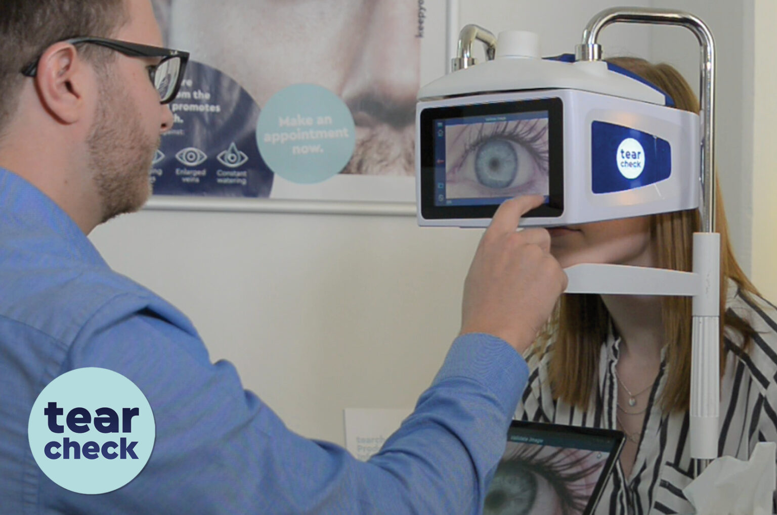
The tearcheck® allows for the visualization of patients’ conditions through two high-resolution cameras that provide fast and clear images and videos with no constraint on the light environment of the examination room. With the second tablet display, the patient can see the results of their exams immediately, supporting an understanding of their overall ocular health and encouraging treatment compliance. Through this easy to use ocular surface visualization device, tearcheck® provides the tools to explain dry eye signs and symptoms by evaluating and identifying dry eye disease as well as MGD.
This attractive and stand-alone analyzer features an intuitive touchscreen user interface, a second tablet device for easy patient-facing display, fully automatic exam analysis, as well as the ability to add new or updated examinations as they are developed in the future in easily downloadable software updates. Since tearcheck® can complete 9 examinations in under 10 minutes time with no constraints on the lighting environment, it is now easy and quick to provide a full dry eye exam on every patient. This allows for the diagnoses of patients with dry eye disease earlier and for the eye care practitioner to recommend individualized treatment protocols based on the in-depth analyses performed. Finally, by seeing the results, the patients have the tools to understand their disease.
9 Examinations Performed in Under 10 Minutes
To begin, the first exam on tearcheck® is the Ocular Surface Disease Index (OSDI) questionnaire, which evaluates the subjective symptoms experienced by the patient. This allows for patient feedback on the severity of their symptoms, giving an overview of their general eye fitness and an indication of their environmental influences. Together with dry eye signs, the OSDI questionnaire is important to quantify dry eye symptoms perceived by the patient to assess several aspects of ocular surface diseases.[4]
The questionnaire is followed by two combined examinations, the Non-Invasive Break-up Time (NIBUT) and the Tear Film Stability Evaluation (TFSE®), both of which use the same imaging, a 10-second video of each eye, as well as the tearcheck® Line Mask, an innovative solution to detect interruptions in the tear film over a wide area on the ocular surface. Here, the NIBUT exam presents the major events appearing on the surface of the tear film by showing where and when the surface of the tear film ruptures, while TFSE® assesses the micro-deformations appearing on the surface of the tear film which reflects instability. The TFSE® is a completely new, patented exam that provides one of the most important diagnostics of dry eye by assessing both the stability of the tear film and showing more finely the surface of the tear film.
The Meibography IR exam visualizes the Meibomian Glands and shows the rate of gland loss in % (compared to an individual with all the glands present) as well as the morphology of the patient’s glands. The real image, the 2D image, and the 3D image of the Meibomian Glands are presented by taking an IR photograph of the inner side of the lower or upper eyelid, which is exposed by using the specially developed tearcheck® Eyelid Flipping Tool, aiding in capturing contact-free images of the glands. IR Meibography is an important diagnostic tool that aids in the identification of patients with MGD,[5] and is especially useful in recognizing the non-obvious form of MGD.[6]
The tearcheck® makes the detection of Demodex possible by examining an enlarged image of the base of the eyelashes, which although does not allow direct visualization of the parasites, makes it possible to find signs of the presence of the mites. The Abortive Blinking exam allows the eye care practitioner to check whether the patient fully closes their eyes when they blink, which is important for the diagnosis of lagophthalmos.
Eye Redness, or conjunctival hyperemia, is detected by displaying the small blood vessels on the ocular surface and lid margins through images with improved contrast. This exam, which shows superficial vascularization of the bulbar and palpebral conjunctiva, allows for the assessment of hyperemia induced by ocular surface inflammation consecutive to dry eye syndromes and also allows the practitioner to follow its evolution over the treatments delivered to the patient.
The final two diagnostic tests, the Ocular Surface Inflammatory Evaluation (OSIE®) and the calculation of Tear Meniscus height both require a drop of fluorescein instilled in each eye before analysis. The completely new patented OSIE® examination is carried out 60 seconds after instilling fluorescein, which is the time necessary for its natural elimination through the tear ducts. Using blue light, areas on the ocular surface that are subject to an increased inflammation risk are detected since fluorescein remains attached to the areas on the ocular surface that are drier. Tear Meniscus height follows the OSIE® examination and uses the residual fluorescein instilled previously, which is present at the level of the lacrimal meniscus, to detect tear meniscus height by quantifying the height of the tear film in the middle of the lower eyelid. The iris is used as the benchmark for tear meniscus height, not the pupil, and so there are no light constraints in the examination room.
The tearcheck® and E>Eye Work Together to Diagnose and Treat Dry Eye Diseases
With Meibomian Gland Dysfunction having been identified as the most common cause of evaporative dry eye disease, having the ability to both accurately diagnose and effectively treat MGD is essential to managing this condition. Used together, tearcheck® provides the necessary dry eye diagnostics and analytical tests necessary for dry eye exploration while the E>Eye device, also Health Canada approved, uses Intense Regulated Pulsed Light (IRPL®) technology to provide a long-lasting solution for the treatment of dry eyes and MGD. While common dry eye management techniques such as gland expression, artificial tears, and warm compresses can have an effect on tear production, taking advantage of new technological advancements and treatment techniques in the area of dry eye disease can have a significant impact on the quality of life of dry eye patients.
[1] Vigo, Luca, Leonardo Taroni, Federico Bernabei, Marco Pellegrini, Stefano Sebastiani, Andrea Mercanti, Nicola Di Stefano, Vincenzo Scorcia, Francesco Carones, and Giuseppe Giannaccare. “Ocular Surface Workup in Patients with Meibomian Gland Dysfunction Treated with Intense Regulated Pulsed Light.” Diagnostics 9, no. 4 (2019): 147. https://doi.org/10.3390/diagnostics9040147.
[2] Korb, Donald R, and Caroline A Blackie. “’Dry Eye’ Is the Wrong Diagnosis for Millions.” Optometry and Vision Science 92, no. 9 (2015): e350–e354. https://doi.org/10.1097/OPX.0000000000000676.
[3] Korb and Blackie, “Dry Eye,” e350-e354.
[4] Guarnieri, Adriano, Elena Carnero, Anne-Marie Bleau, Belen Alfonso-Bartolozzi, and Javier Moreno-Montañés. “Relationship Between OSDI Questionnaire and Ocular Surface Changes in Glaucomatous Patients.” International Ophthalmology 40 no. 3 (December 2019): 741–751. https://doi.org/10.1007/s10792-019-01236-z.
[5] Srinivasan, Sruthi, Kara Menzies, Luigina Sorbara, and Lyndon Jones. “Infrared Imaging of Meibomian Gland Structure Using a Novel Keratograph.” Optometry and Vision Science 89, no. 5 (May 2012): 788–794. https://doi.org/10.1097/opx.0b013e318253de93.
[6] Pult, Heiko and Britta H Riede‐Pult. “Non‐Contact Meibography in Diagnosis and Treatment of Non‐Obvious Meibomian Gland Dysfunction.” Journal of Optometry 5, no. 1 (2012): 2–5. https://doi.org/10.1016/j.optom.2012.02.003.